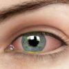
Keratoconus is a condition that affects the cornea – the front surface of the eye – to thin and bulge, creating a “cone” like feature. This condition may begin to occur during puberty and progressively becomes worse over time. Blurry vision is a major symptom for keratoconus, with many patients reporting “hazing” or a second image on top of the first. Eye rubbing, a history of affected family members with the same condition, eczema, or a history of refractive surgery might also put someone at more risk of keratoconus.
When managed properly by an optometrist from an early stage, this disease will not interfere with daily life and activity. Once a diagnosis is made by your optometrist, the patient will be referred to see an ophthalmologist for further assessment. There are a variety of treatment options – both surgical and non-surgical – that an optometrist and ophthalmologist can offer to help alleviate the symptoms of keratoconus.
NON-SURGICAL
At Eyecare Network, your optometrist can offer contact lenses which are the hallmark for managing this disease. There are multiple types that your optometrist can offer, including:
- Custom soft contact lenses
- Rigid gas permeable (RGP)
- Hybrid contact lenses (hard centre and a soft skirt)
- Scleral contact lenses
- Piggyback contact lenses (soft lens on top of a RGP)
To fit these types of lenses, complex imaging technology of the cornea is required to get good results. Eyecare Network is well equipped with a Medmont Corneal Topographer Anterior ocualar coherence topographer help with accurate contact lens fittings.
SURGICAL
At Eyecare Network, you may also have a discussion with your optometrist about surgical options, if they are right for you.
- Surgical management of keratoconus may include a collagen cross linking procedure. This can be a very good preventative option to strengthen the fibres in the cornea to prevent the progression of keratoconus. In this procedure, vitamin B is instilled into the eye which is then exposed to UV radiation to help form links between the fibres of the cornea.
- Corneal INTACS is also another procedure that helps reshape the bulging cornea, with the insertion of two semi-circular rings into the cornea. INTACS help to flatten the centre of the cornea for patient comfort and better vision.
- For advanced cases where the damage cannot be managed by other treatment options, a patient may undergo a corneal graft. This is a highly successful procedure (90-97% success) where a partial or full thickness graft transplant may occur, depending on the circumstance.
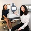
To discuss further options or to find out more, book in to see one of our highly knowledgeable optometrists and friendly staff on 9728 7288.
HOW IS KERATOCONUS DIAGNOSED BY MY OPTOMETRIST?
A full ocular examination of the health of your eye can reveal some telltale signs of keratoconus.
- A biomicroscope/slit lamp is used to examine the cone with a magnified view as well as the different layers of the cornea. We look at the base of the cone and the apex, also for any thinning in the front few layers of the eye.
- A corneal topographer to examine the shape and features of the surface of the cornea.
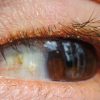
- Examination with the naked eye is also possible where in advanced cases of keratoconus, the bulging of the lower lid in downgaze (Munson’s sign) is observable.